26 May Looking at the eyes of Alzheimer’s Disease
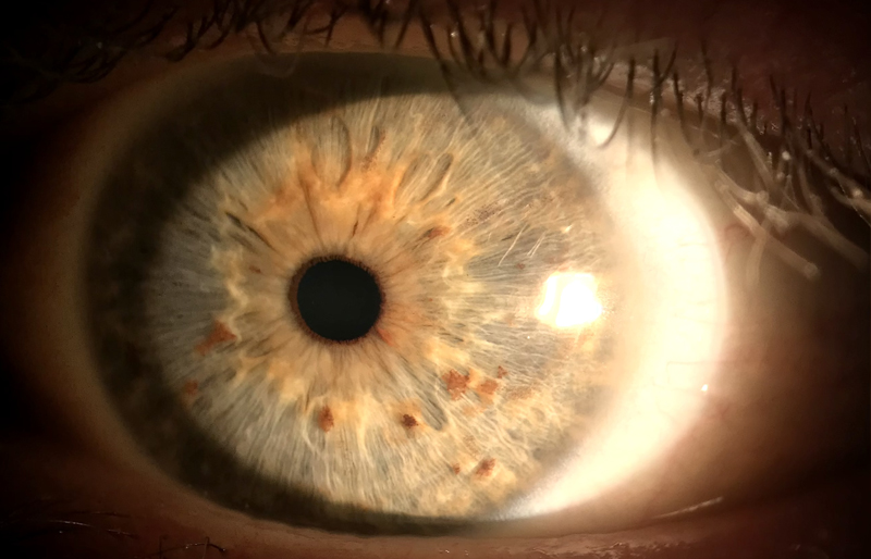
The retina is a brain tissue with a very special quality that makes it unique amongst other brain tissues. It is located in the back of the eye and everything in front of it is transparent in order to allow light to pass through. This transparency is used by ophthalmologists to visually access the retina (we are able to see neural tissue directly by just observing the back of the eye) and thanks to modern technology this technique has gradually gained more detail.

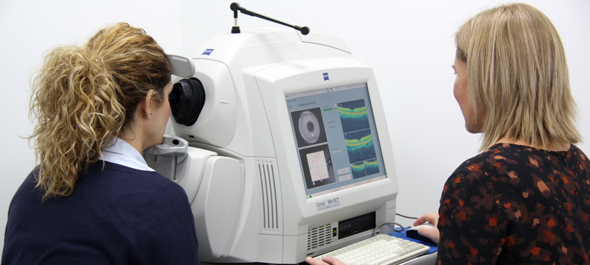
Eye check-ups can detect other conditions that are not related to the eye. Visioker Eye Clinic Torrevieja
It is not strange to consider that when the brain is affected by other diseases there would also be some effect on the retina, since it is brain tissue. If we could know what changes happen in the retina (by observing it) we would be able to diagnose neurological diseases and how they evolve.
Investigators have found that patients with Alzheimer’s disease (AD) also show some characteristics in their optic nerve. This study started at the end of the last century, and more recently this concept has acquired great relevance due to the Retinal Optical Coherence Tomography or OCT. An OCT is an imaging technique used in ophthalmology for advanced diagnosis of retinal and optic nerve pathologies. It is based on optical interferometry and allows us a detailed view of the retina by studying each of its layers and analyzing thicknesses in different areas. The OCT gave way to another technology called OCT- Angiography or OCTA which bases itself on the movement of red blood cells in the vessels and allows us to observe and quantify in detail the retinal vasculature. (It is like an angiography without the need of using dye contrasts.)
Structural OCT and OCTA are both noninvasive imaging techniques and they can take as many images as necessary. Thus, the interest to see if we can find differential markers among healthy patients and AD patients that appear in early stages of the disease. This would also be of great help regarding investigation as it would allow us to monitor the evolution of the disease and test different drugs in early stages.

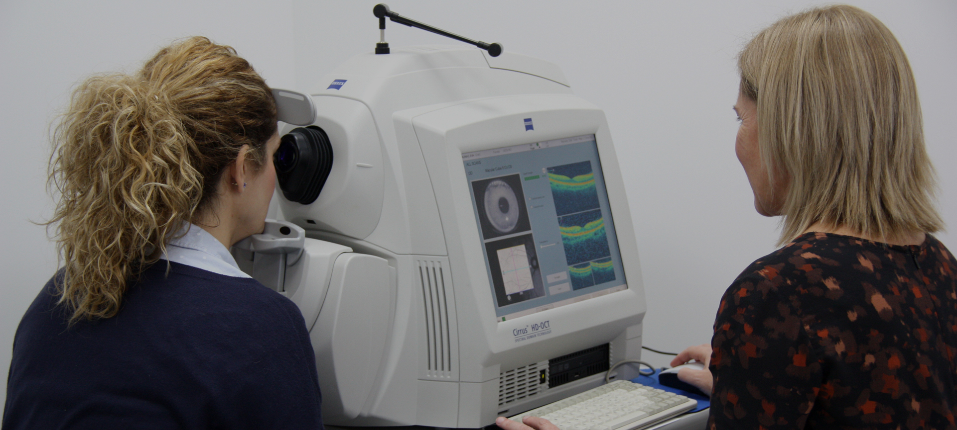
OCT and OCTA imaging at Visionker – Eye Clinic Torrevieja
Numerous structural OCT and OCTA studies reveal evident changes in AD patients in relation to healthy patients.
- Retinal nerve fiber layer (RNFL) thinning, especially in the superior and inferior quadrants.
- Retinal ganglion cell layer and inner plexiform layer (GCL + IPL) degeneration.
- Macular volume reduction.
- Macular perfusion (blood flow) reduction.
However, the limits between healthy patients and affected patients are still not clear to apply on everyday clinical work. Some of these changes can be seen in other neurodegenerative diseases such as Parkinson or Huntington disease. There is still a lot to achieve but, currently there are several studies in place trying to pinpoint these changes and use them as markers.
Some studies have achieved to detect retinal amyloid β deposits in these patients, which is very specific to Alzheimer’s disease. The exploration was done with a confocal laser looking at the superior quadrants of the retina before and after administering curcumin (fluorescent linked to the amyloid β deposits).
We can therefore confirm that the near future is full of hope for improving on early detection of Alzheimer’s disease. This will allow studies on drugs that act in early stages of AD. Ophthalmology can help with this objective and, in addition to brain PET scans and MRIs, we have seen how it is possible to contribute noninvasively in an early detection of AD looking at the eyes of Alzheimer’s disease. Perhaps technology and artificial intelligence will be the key to early detection of this horrible disease and help find an effective solution to its prevention.
Dr. Eva M. Salinas Martínez, Eye Doctor at Visionker Eye Clinic, Torrevieja
Dr. José I. Belda Sanchis, Eye Doctor at Visionker Eye Clinic, Torrevieja
IF YOU WANT TO LEARN MORE:
Cunha LP, Lopes LC, Costa-Cunha LVF, Costa CF, Pires LA, Almeida ALM, et al. Macular Thickness Measurements with Frequency Domain-OCT for Quantification of Retinal Neural Loss and its Correlation with Cognitive Impairment in Alzheimer’s Disease. PLoS ONE. 2016;11(4):e0153830.
Altered macular microvasculature in mild cognitive impairment and Alzheimer disease [Internet]. Disponible en: https://www.ncbi.nlm.nih.gov/pmc/articles/PMC5902666/
Koronyo Y, Biggs D, Barron E, Boyer DS, Pearlman JA, Au WJ, et al. Retinal amyloid pathology and proof-of-concept imaging trial in Alzheimer’s disease. JCI Insight [Internet]. 17 de agosto de 2017 ;2(16). Disponible en: https://www.ncbi.nlm.nih.gov/pmc/articles/PMC5621887/
AFAE – Asociación de Familiares de Personas con Alzheimer de Elche, pueden encontrar ayuda e información en esta asociación.


Sorry, the comment form is closed at this time.